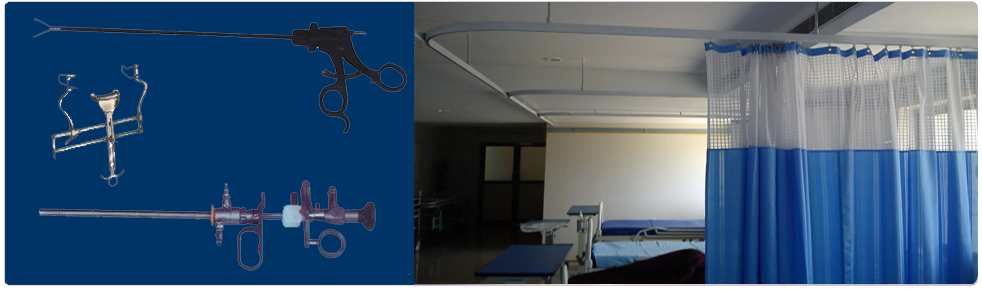
Surgical Instruments

Retractors Many varieties and sizes of retractors are available, and the use of specific retractors will largely depend on the type of surgical procedure being performed. Retractors are used for holding the incision open to provide exposure to the surgical site. Smaller types held by the fingers or hand retract skin and subcutaneous tissue in shallow surgical areas. Larger, heavier models retract muscle tissue and organs in deeper surgical sites. Some retractors are held in place by an assistant while the surgeon completes the procedure, while self-retaining retractors require no assistant to hold them. Self-retaining retractors are held open by their own action and may be used in conjunction with the hand held retractors. Examples of retractors: RichardsonEastman, Mayo, Jansen Mastoid, Weitlaner, Cerebellum, Gelpi, Volkman Rake, Green Goiter, Army-Navy, Deaver.

Laparascopic/Endoscopic

| Technical Specification | Hfease100 | |
| Max Power | 400W | |
| Display | 7 Segment LED Display | |
| Program | 99 | |
| Monopolar Cut Modes | 2 Cut, 2 Blend, Endo Cut | |
| Auto Stop Coagulation Modes | Present | |
| Monopolar Coagulation Modes | Spray, Dessicate, Pulgurate, Soft | |
| Auto Stop Coagulation in Bipolar | >=20/>=15 | |
| Bipolar Modes | Cut, Force, Micro | |
| Tissue Response Circuit | Present | |
| Patient Plate Monitoring System | Present | |
| Dimension ( W x D x H ) | 360mm x 145mm x 305mm | |
| Technical Specification | ||
| Model | ENCO2-20H | ENCO2-30H |
| Insufflation Pressure | 4 - 30 mm Hg | 0 - 30 mm Hg |
| Dimension(WxHxD) | 210mm x 80mm x 230mm | 290mm x 105mm x 260mm |
| Weight | 2 kg | 2.5 kg |
| Auto Stop Coagulation Modes | 2 kg | 2.5 kg |
| Gas | C02 (Medical) | C02 (Medical) |
| Gas Connection | UNF 7/16" | UNF 7/16" |
| Power Consumption | 15 Watts Maximum | 15 Watts Maximum |
| Input Pressure Minimum | 3 Bar | 1 Bar |
| Input Pressure Maximum | 10 Bar | 10 Bar |
| Output Connection | 4 to 5mm ID tube | 4 to 5mm ID tube |
| Output Connection | 4 to 5mm ID tube | 4 to 5mm ID tube |
| Flow Rate Setting | 1 to 20 Ltr/min | 1 to 30 Ltr/min |
| Keyboard | Feather Touch | Feather Touch |
| Gas Warmer | Builtin | Builtin |
| Keyboard | Feather Touch | Feather Touch |
| Manufactured and tested | IEC 601-1 / EN 60601-1 | IEC 601-1 / EN 60601-1 |
| Input Supply Voltage | 230 VAC + 15% @ 50/60 Hz (110 V optional) | 230 VAC + 15% @ 50/60 Hz (110 V optional) |


Laparoscopy is a surgery that uses a thin, lighted tube put through a cut (incision) in the belly to look at the abdominal organs or the female pelvic organs.Laparoscopy is used to find problems such as cysts, adhesions, fibroids , and infection. Tissue samples can be taken for biopsy through the tube (laparoscope).
In many cases laparoscopy can be done instead of laparotomy surgery that uses a larger incision in the belly. Laparoscopy can be less stressful and may have less problems and lower costs than laparotomy for minor surgeries. It can often be done without needing to stay overnight in the hospital.
A laparoscope is a small tube that consists of a light source and a camera. The camera relays images of the inside of the abdomen or pelvis to a television monitor.
The surgeon makes a small incision in the skin and passes the laparoscope through it to study the organs and tissues inside the abdomen or pelvis.
The advantages of this technique over traditional open surgery are that people who have a laparoscopy have:
• less pain after the operation
• minimal scarring
While laparoscopic surgery to completely remove cancerous tumors, surrounding tissues, and lymph nodes is used on a limited basis, this type of operation is widely used in noncancerous conditions that once required open surgery.
Laparoscopy can be used in determining the spread of certain cancers. Sometimes it is combined with ultrasound. Although laparoscopy is a useful staging tool, its use depends on a variety of factors, which are considered for each patient. Laparoscopy is sometimes used as part of a palliative cancer treatment. This type of treatment is not a cure, but can often lessen the symptoms. An example is the feeding tube, which cancer patients may have if they are unable to take in food by mouth. The feeding tube provides nutrition directly into the stomach. Inserting the tube with a laparoscopy saves the patient the ordeal of open surgery.


Laparoscopic surgery requires only a small incision and is commonly used today for appendectomies, hysterectomies, and prostatectomies. Patients lose much less blood during and after surgery and recover much faster, compared to other surgical procedures.
As with any surgery, patients should notify their physician of any medications they are taking (prescription, over-the-counter, or herbal) and of any allergies. Precautions vary due to the several different purposes for laparoscopy. Patients should expect to rest for several days after the procedure, and should set up a comfortable environment in their home (with items such as pain medication, heating pads, feminine products, comfortable clothing, and food readily accessible) prior to surgery.
A gastrointestinal endoscope may be inserted through the mouth or anus. An ultrasound probe can be added to a gastrointestinal endoscope. This is called an endoscopic ultrasound. Depending on the area of interest, this device can also be passed through the mouth or anus.
An endoscopy is often used to confirm a diagnosis when other devices, such as an MRI, X-ray, or CT scan are considered inappropriate.
An endoscopy is often carried out to find out the degree of problems a known condition may have caused. The endoscopy, in these cases, may significantly contribute towards the doctor's decision on the best treatment for the patient.
